بررسی شبکیه

Oct Macula

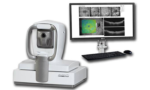
OCT برای ارزیابی دقیق اختلالات ماکولا بسیار ارزشمند است. قبل و بعد از عمل جراحی کاتاراکت انجام این تست ضروری است؛ چرا که پیش آگهی وضع بیمار قبل از عمل لازم است و ضمنا برای بررسی علل ناکامل بودن دید بیمار یک هفته بعد از عمل کمک می کند تا از وضعیت ماکولا مطمئن شویم چرا که از اختلالات لایه های مختلف رتین و کوروئید که ممکن است در دید بیمار تاثیر داشته باشد ما را مطلع می سازد.

با امتحان کلینیکی ته چشم فقط 28% Lamellar Hole که با OCT مشخص شده را می توان تشخیص داد. باید توجه داشت که اکثر ویتریورتینال ترکشن و مامبران اپی رتینال بدون این وسیله تشخیص داده نمی شود و ضمنا باید دانست که جراحی کاتاراکت اکثرا چنین اختلالاتی را به وجود نمی آورند بلکه از قبل زمینه آن فراهم بوده است.

پاتولوژیهای رتین مثل ویتریوما کولرترکشن،لاملار ماکولار Hole و آترئفی کوروییدسنیلرا اکثرا در امتحان ته چشم نمی توان تشخیص داد.

با OCT هیالویید خلقی و ارتباط آنرا با ماکولا می توان تشخیص داد ناحیه ای که در ماکولا ادما و دژسانس ماکولا رل عمده دارد و در واقع تنها وسیله ای است که اختلالات کوروئید را دقیقا مشخص می کند.

آتروفی کوروئید یک عامل خطرساز برای گلوکوم به شمار می آید مخصوصا وقتی همراه با آتروفی پری پاپیلار باشد. این بیماران دید خوبی نخواهند داشت حتی اگر ماکولا سالم به نظر می رسد.

دژنرسانس کوریورتینال میوپیک نیز همراه با نقصان بینایی ممکن است باشد.

گاه متعاقب جراحی کاتاراکت دژنرسانس ویتره ممکن است ایجاد شده و یا تشدید شده و سبب جداشدگی خلفی ویتره شود و این عارضه به نوبه خود سبب ویتریوماکولار ترکشن و یا پیشرفت مامبران اپی ماکولار و تشدید علائم بشود و یا حتی ماکولار Hole ایجاد نماید. یک هفته بعد از جراحی کاتاراکت 20% این بیماران دچار جداشدگی خلفی می شوند.




بازگشت به لیست
Retinal examination

Oct Macula

In order to accurately evaluating macular degeneration, the OCT is highly valuable. This test is necessary before and after cataract surgery, because a pre-knowledge of the patient’s condition is a must before the operation, moreover, it helps to make sure of the macular condition in investigations on the causes of patient’s incomplete vision one week after surgery because it informs us about the disorders of the various retina and choroid layers that may affect the patient’s vision.

Through clinical examination of the eye bottom, only 28% of the lamellar hole was detected by the specified OCT. It should be noted that most of the Vitreoretinal Traction and Epiretinal Membranes are not detected without this device, and it should also be noted that cataract surgery often does not cause such disorders, but the grounds were already provided.

Retinal pathologies, such as vitreomacular traction, lamellar macular hole, and Senile Choroidal Atrophy are most often not detectable in bottom eye examination.

With OCT, mood hyaloid and its relation with macula we can detect that a region has a major role in macular edema and macular degeneration, and is in fact the only means that accurately identifies Choroidal disorders.

Choroidal atrophy is a risk factor for glaucoma, especially when it is accompanied by parapapillary atrophy. These patients will lack a clear vision, even if the macula seems healthy.

myopic chorioretinal degeneration may also be associated with visual impairment.

Sometimes, after cataract surgery, vitrea degeneration may develop or exacerbate and posterior vitreous detachment, which in turn, causes vitreomacular traction or Epimacular membrane progression and exacerbation of symptoms, or even cause macular hole. One week after cataract surgery, 20% of these patients suffered with posterior detachment.


بازگشت به لیست